Home

strâmt Frizer Port red microscopic light pantă câteva Funcționare posibilă

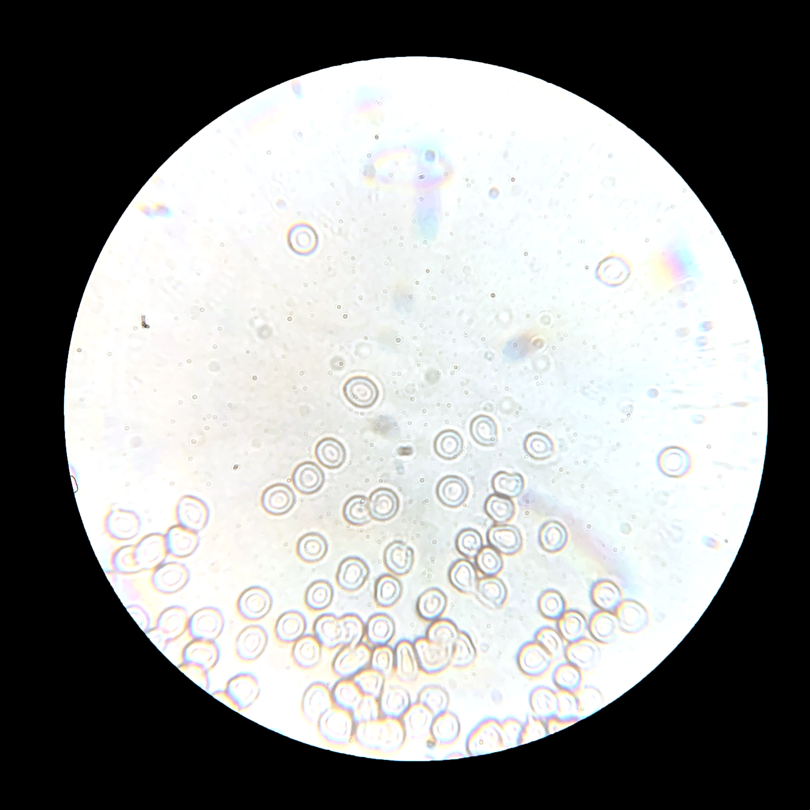
Scientific Image - Human Red Blood Cells | NISE Network

Light microscopy images of exerciseinduced changes in red blood cells... |  Download Scientific Diagram
Light microscopy images of exerciseinduced changes in red blood cells... | Download Scientific Diagram

Under the Microscope: Blood | Office for Science and Society - McGill  University
Under the Microscope: Blood | Office for Science and Society - McGill University

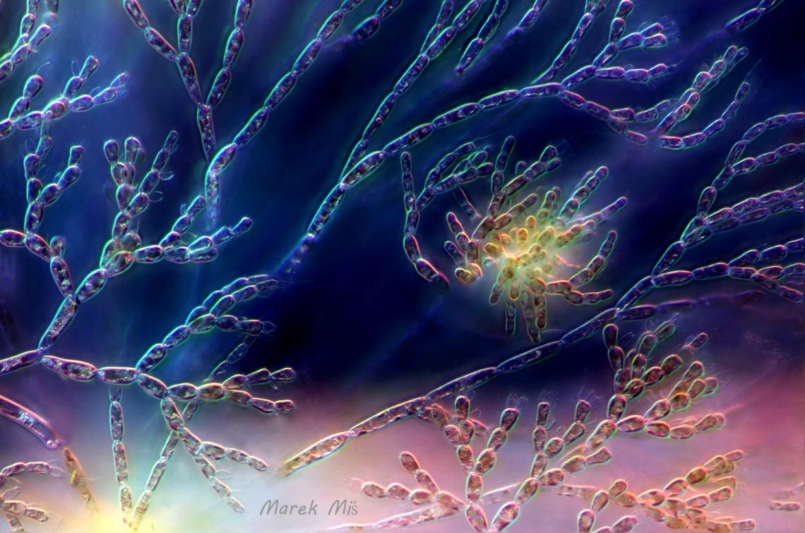
Red Alga Under the Microscope Microscope Art Microscopic - Etsy

Red blood cell morphology by light microscopy. a Control (without... |  Download Scientific Diagram
Red blood cell morphology by light microscopy. a Control (without... | Download Scientific Diagram

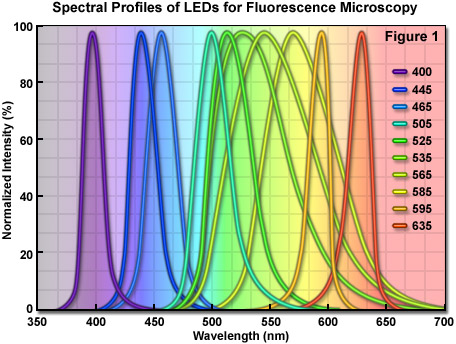
ZEISS Microscopy Online Campus | Light-Emitting Diodes

Choosing a ring light for your microscope | Pathwooded

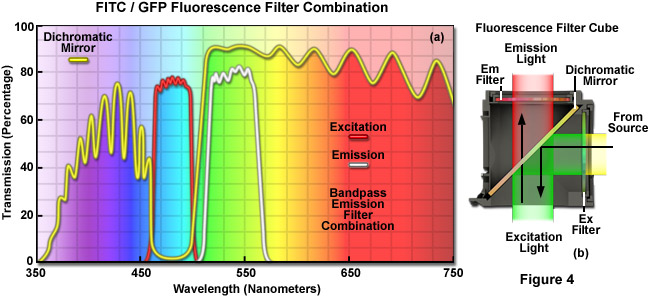
ZEISS Microscopy Online Campus | Microscopy Basics | Fluorescence Microscopy

Morphology of red blood cells stained on day a) 0, b) 5, c) 10, d) 15... |  Download Scientific Diagram
Morphology of red blood cells stained on day a) 0, b) 5, c) 10, d) 15... | Download Scientific Diagram

Amyloid on Congo red: red on light microscopy (A) and apple-green under...  | Download Scientific Diagram
Amyloid on Congo red: red on light microscopy (A) and apple-green under... | Download Scientific Diagram

Three human parasite form patterns, trophozoite form of Plasmodium malariae  malaria infected red blood cells on thin film blood smear under 100X light  microscope (Selective focus). Stock Photo | Adobe Stock
Three human parasite form patterns, trophozoite form of Plasmodium malariae malaria infected red blood cells on thin film blood smear under 100X light microscope (Selective focus). Stock Photo | Adobe Stock
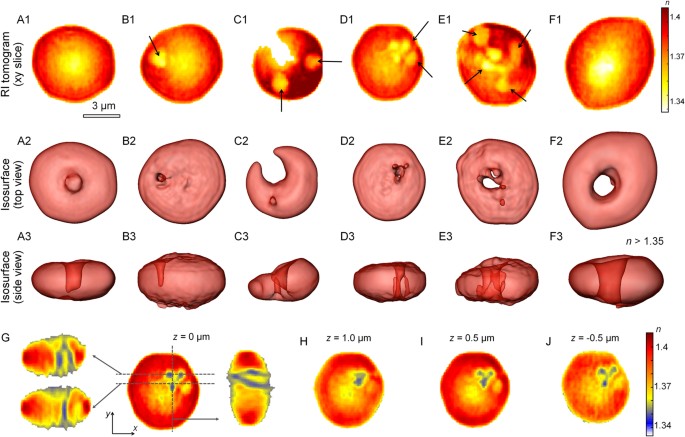
Characterizations of individual mouse red blood cells parasitized by  Babesia microti using 3-D holographic microscopy | Scientific Reports
Characterizations of individual mouse red blood cells parasitized by Babesia microti using 3-D holographic microscopy | Scientific Reports
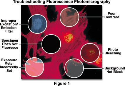
Fluorescence Microscopy Errors | Olympus LS

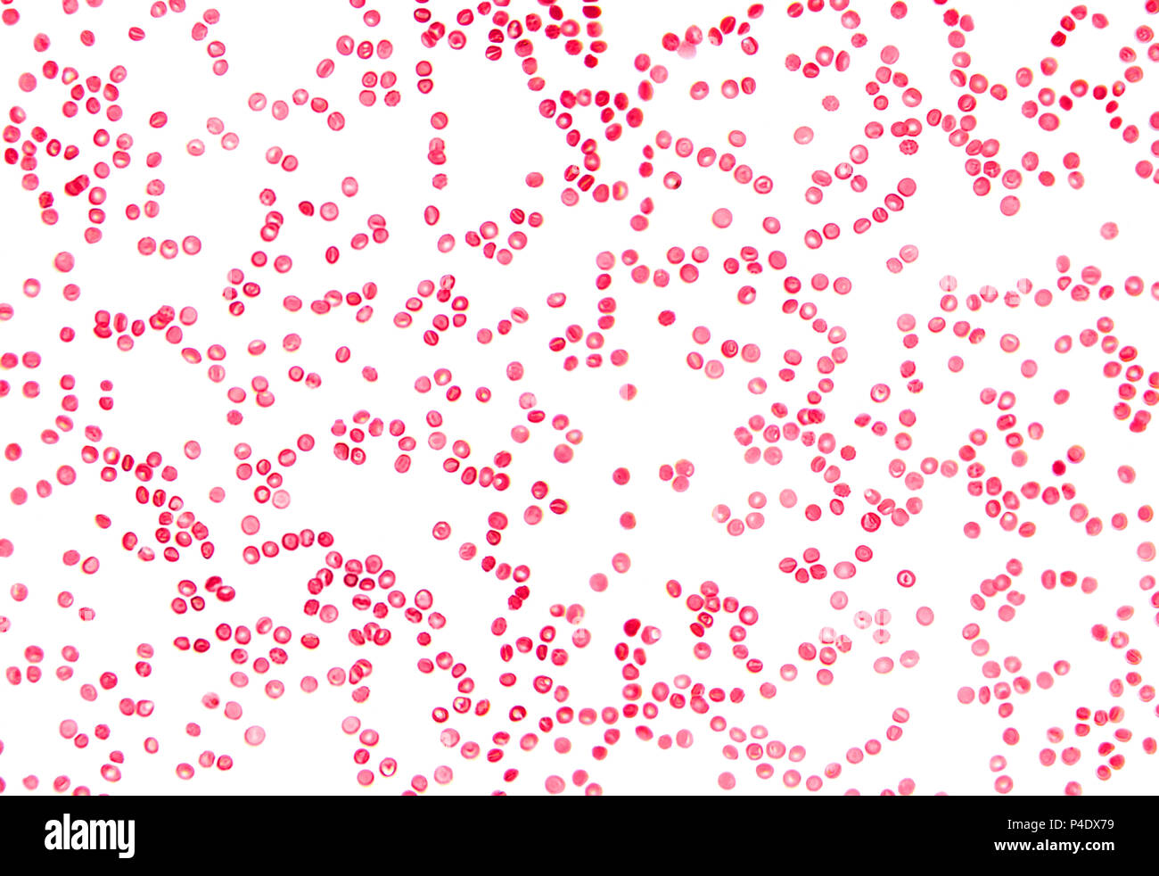
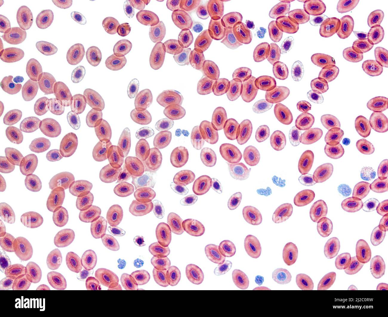
Light micrograph red blood cells hi-res stock photography and images - Alamy

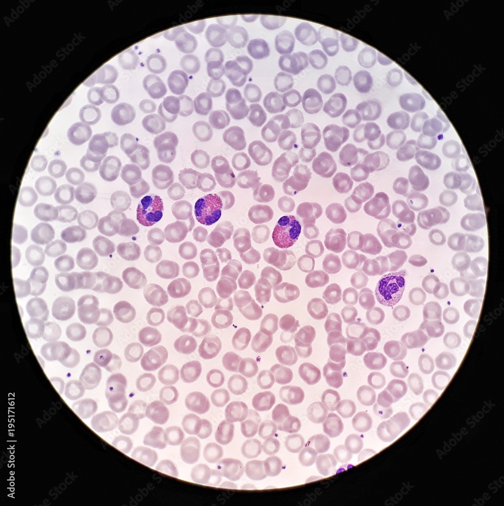
Human blood smear under 100X light microscope with Eosinophils, Neutrophil  and hypochromic red blood cells (Selective focus). Stock Photo | Adobe Stock
Human blood smear under 100X light microscope with Eosinophils, Neutrophil and hypochromic red blood cells (Selective focus). Stock Photo | Adobe Stock

Ways to Examine Metals by Light Microscopy - YouTube

Blood vessels with red blood cells, transevrse section, light micrograph,  photo under microscope, Stock Photo, Picture And Low Budget Royalty Free  Image. Pic. ESY-052883581 | agefotostock
Blood vessels with red blood cells, transevrse section, light micrograph, photo under microscope, Stock Photo, Picture And Low Budget Royalty Free Image. Pic. ESY-052883581 | agefotostock
Red blood cells, light microscopy - Stock Video Clip - K004/5822 - Science  Photo Library
Red blood cells, light microscopy - Stock Video Clip - K004/5822 - Science Photo Library

Scanning Electron Microscopy of the Human Cornea

21,224 Red Cells Microscope Images, Stock Photos & Vectors | Shutterstock

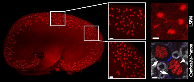
Light Sheet Microscopy: acquire 3D quantitative images of whole organs with  cellular resolution | Light & Electron Microscopy for Biology
Light Sheet Microscopy: acquire 3D quantitative images of whole organs with cellular resolution | Light & Electron Microscopy for Biology
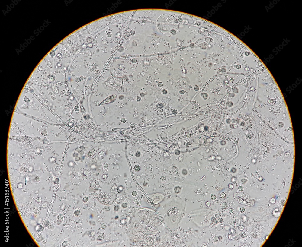
Abnormal result of urinalysis examination from microscopic method under 40X light  microscope; show many white blood cells (WBC), red blood cells (RBC),  epithelial cells, bacteria and hyphae of fungus. Stock Photo
Abnormal result of urinalysis examination from microscopic method under 40X light microscope; show many white blood cells (WBC), red blood cells (RBC), epithelial cells, bacteria and hyphae of fungus. Stock Photo

Light microscope observations of untreated human red blood cells... |  Download Scientific Diagram
Light microscope observations of untreated human red blood cells... | Download Scientific Diagram

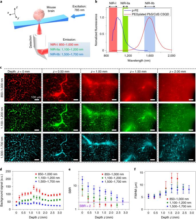
Light-sheet microscopy in the near-infrared II window | Nature Methods

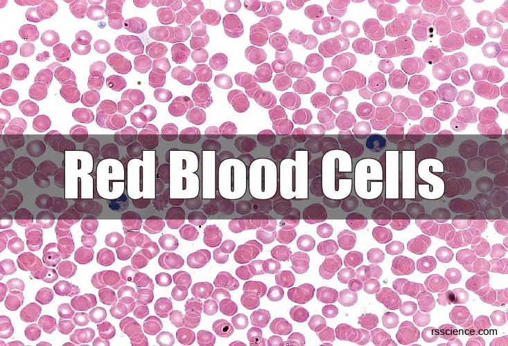
Red Blood Cells – Definition, Biology, and Observation under the Microscope

Molecular Expressions Microscopy Primer: Light and Color - Diffraction of  Light
Molecular Expressions Microscopy Primer: Light and Color - Diffraction of Light

Light micrograph red blood cells hi-res stock photography and images - Alamy