Home

snorkel broderie Aparține fluorescence microscope purkinje neuron radar Charles Keasing comerț

Complex brain cell connections in the cerebellum more common than believed

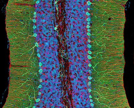
Purkinje Nerve Cells In The Cerebellum Metal Print by Thomas Deerinck,  Ncmir - Fine Art America
Purkinje Nerve Cells In The Cerebellum Metal Print by Thomas Deerinck, Ncmir - Fine Art America

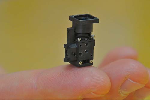
Scientists Develop Miniaturized Fluorescence Microscope for use in Live  Brain Imaging, Parallel Screening and Portable Diagnostics
Scientists Develop Miniaturized Fluorescence Microscope for use in Live Brain Imaging, Parallel Screening and Portable Diagnostics
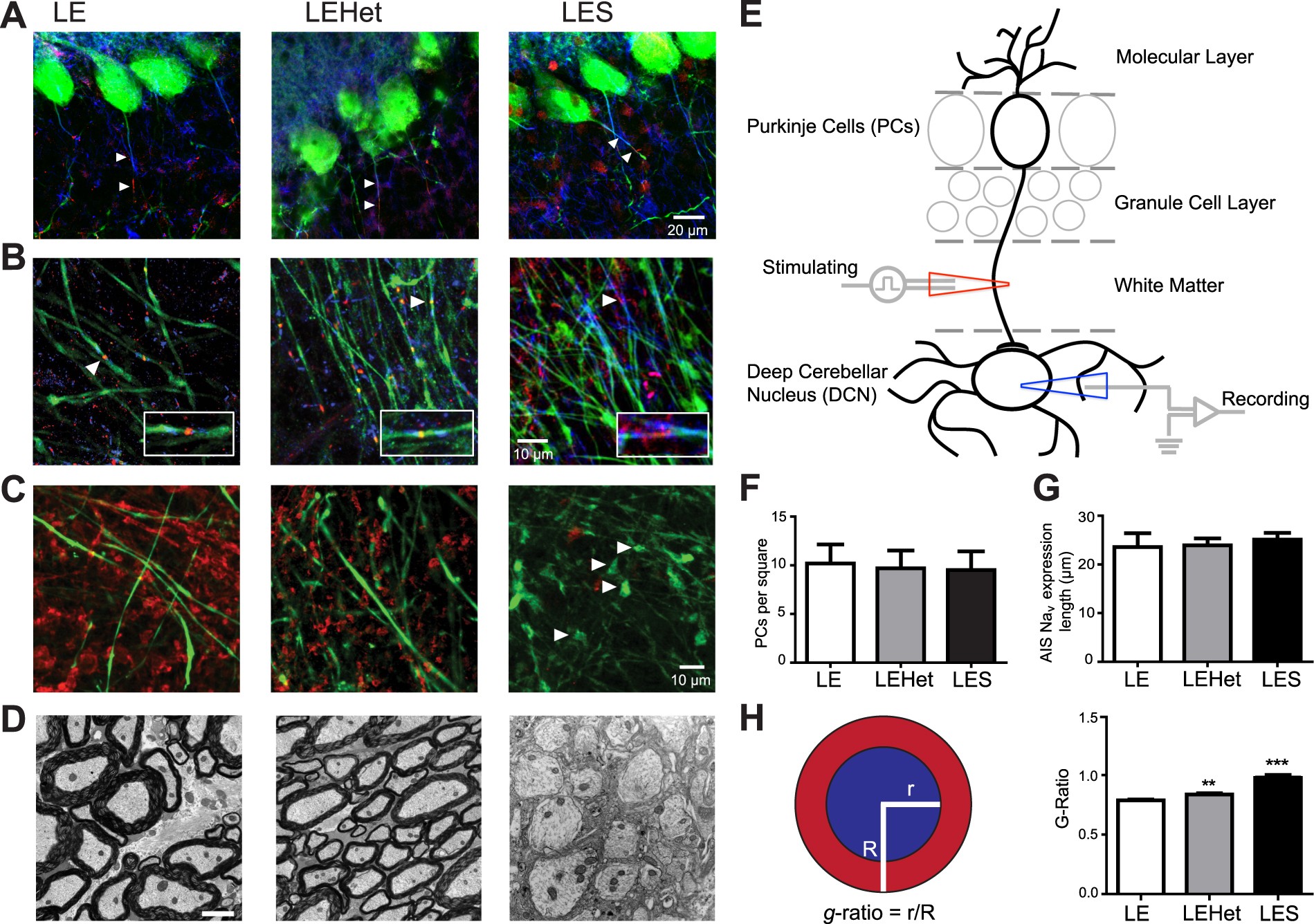
Myelination of Purkinje axons is critical for resilient synaptic  transmission in the deep cerebellar nucleus | Scientific Reports
Myelination of Purkinje axons is critical for resilient synaptic transmission in the deep cerebellar nucleus | Scientific Reports
Direct reprogramming of cardiomyocytes into cardiac Purkinje-like cells -  ScienceDirect
Direct reprogramming of cardiomyocytes into cardiac Purkinje-like cells - ScienceDirect

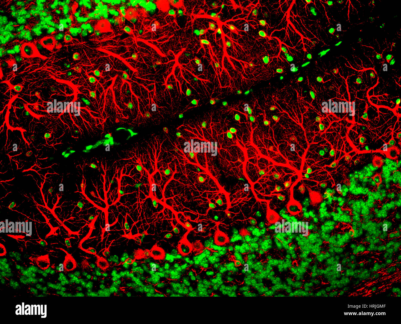
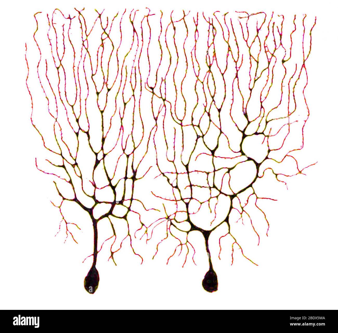
Purkinje Cells, Fluorescent LM Stock Photo - Alamy

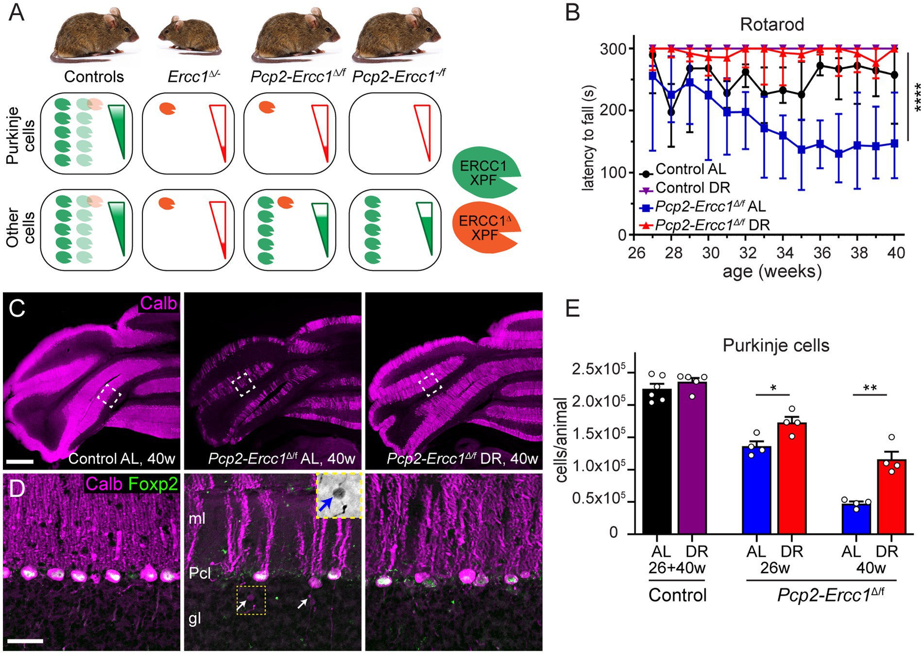
Frontiers | Purkinje-cell-specific DNA repair-deficient mice reveal that  dietary restriction protects neurons by cell-intrinsic preservation of  genomic health
Frontiers | Purkinje-cell-specific DNA repair-deficient mice reveal that dietary restriction protects neurons by cell-intrinsic preservation of genomic health

Mouse Purkinje (brain) cells | Purkinje Cell | Nikon Small World |  Microscopic photography, Neurons, Brain art
Mouse Purkinje (brain) cells | Purkinje Cell | Nikon Small World | Microscopic photography, Neurons, Brain art
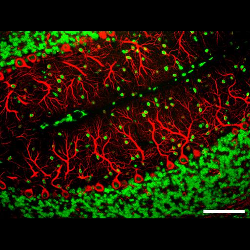
CIL:34544, Mus musculus, CNS neuron (sensu Vertebrata), Purkinje cell. CIL.  Dataset
CIL:34544, Mus musculus, CNS neuron (sensu Vertebrata), Purkinje cell. CIL. Dataset

Maximise Data Return with Confocal Microscopy - BioTechniques

Purkinje nerve cell, light micrograph - Stock Image - C052/2323 - Science  Photo Library
Purkinje nerve cell, light micrograph - Stock Image - C052/2323 - Science Photo Library

Purkinje nerve cell, confocal micrograph - Stock Image - C039/7457 -  Science Photo Library
Purkinje nerve cell, confocal micrograph - Stock Image - C039/7457 - Science Photo Library

Fluorescent Dyes | Science Lab | Leica Microsystems

Filling a Purkinje neuron with fluorescent dye | Okinawa Institute of  Science and Technology OIST
Filling a Purkinje neuron with fluorescent dye | Okinawa Institute of Science and Technology OIST

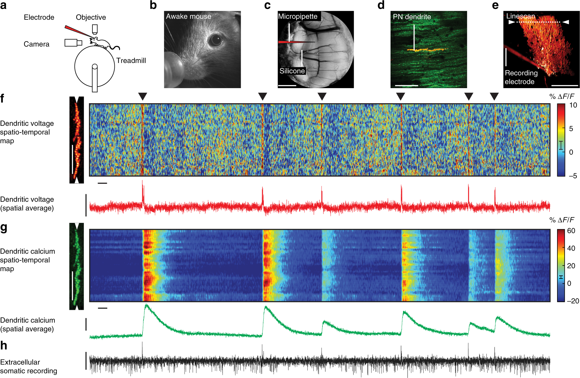
Simultaneous dendritic voltage and calcium imaging and somatic recording  from Purkinje neurons in awake mice | Nature Communications
Simultaneous dendritic voltage and calcium imaging and somatic recording from Purkinje neurons in awake mice | Nature Communications

Automated segmentation of brain cells for clonal analyses in fluorescence  microscopy images - ScienceDirect
Automated segmentation of brain cells for clonal analyses in fluorescence microscopy images - ScienceDirect

Purkinje Cell — Cells of Life

CIL:40124, Mus musculus, Purkinje cell. CIL. Dataset

Electron microscopy of normal and degenerating Purkinje cell bodies and...  | Download Scientific Diagram
Electron microscopy of normal and degenerating Purkinje cell bodies and... | Download Scientific Diagram
Purkinje nerve cells cerebellum hi-res stock photography and images - Alamy

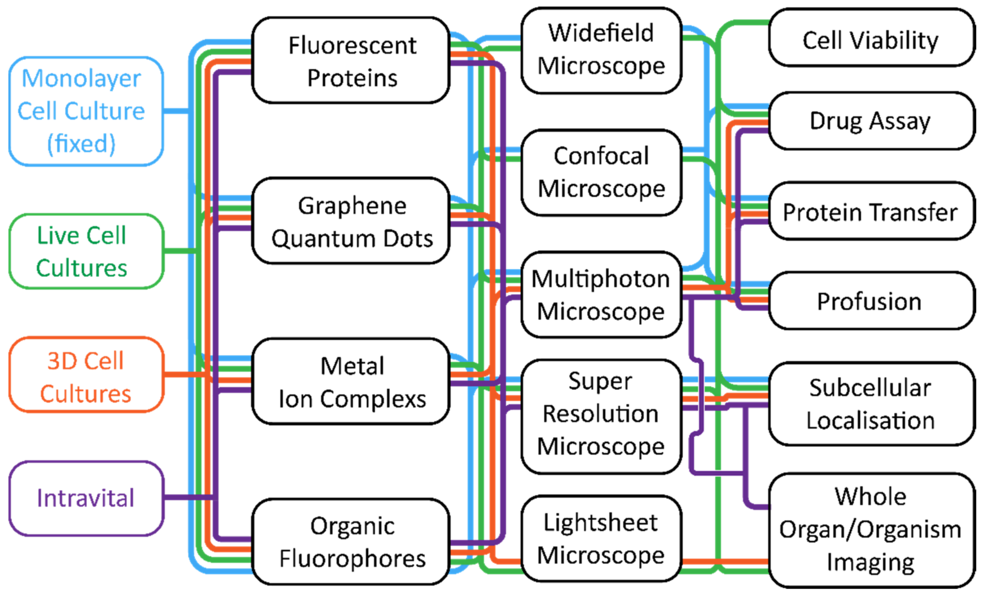
Cells | Free Full-Text | Fluorescence Microscopy—An Outline of  Hardware, Biological Handling, and Fluorophore Considerations
Cells | Free Full-Text | Fluorescence Microscopy—An Outline of Hardware, Biological Handling, and Fluorophore Considerations

eLife - the journal on X: "Scientists have developed a two-photon fluorescence  microscope coupled with adaptive optics, allowing in vivo imaging of the  mouse retinal structure and function. https://t.co/nZqlmUOQIy  https://t.co/wAQ8xsJgNo" / X
eLife - the journal on X: "Scientists have developed a two-photon fluorescence microscope coupled with adaptive optics, allowing in vivo imaging of the mouse retinal structure and function. https://t.co/nZqlmUOQIy https://t.co/wAQ8xsJgNo" / X

CIL:41465, Rattus, glial cell, purkinje neuron | Library Digital  Collections | UC San Diego Library
CIL:41465, Rattus, glial cell, purkinje neuron | Library Digital Collections | UC San Diego Library

Microglia Promote the Death of Developing Purkinje Cells: Neuron

Art of Science Competition / Gallery - Purkinje Neurons